⚠️ Disclaimer: The information on this page is intended for general educational purposes only and does not constitute medical advice. It is not a substitute for professional medical diagnosis, treatment, or guidance. Always consult a qualified healthcare provider before beginning any exercise or rehabilitation program.
Musculoskeletal Health
Shoulder Pain
Explore causes, diagnosis, and step-by-step rehab strategies to restore shoulder strength and confidence.
Start Here
Understanding Shoulder Pain Basics
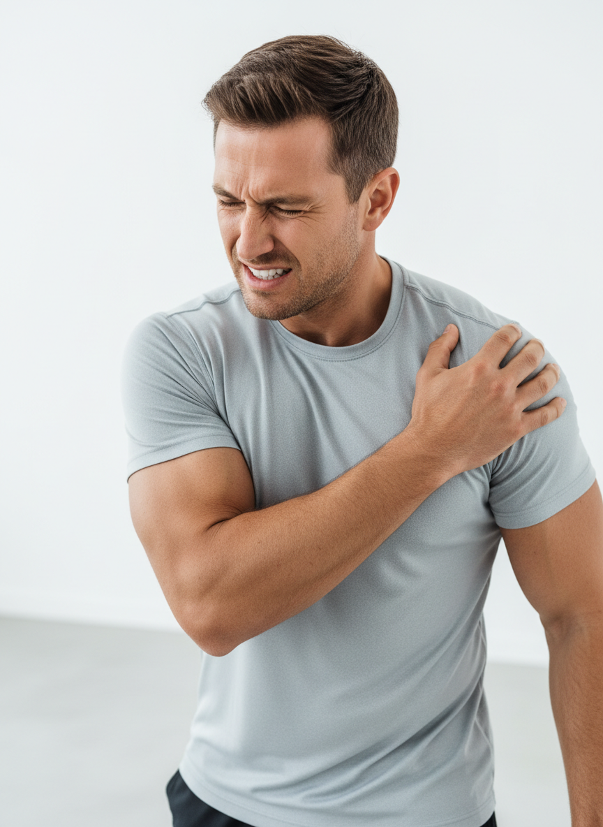
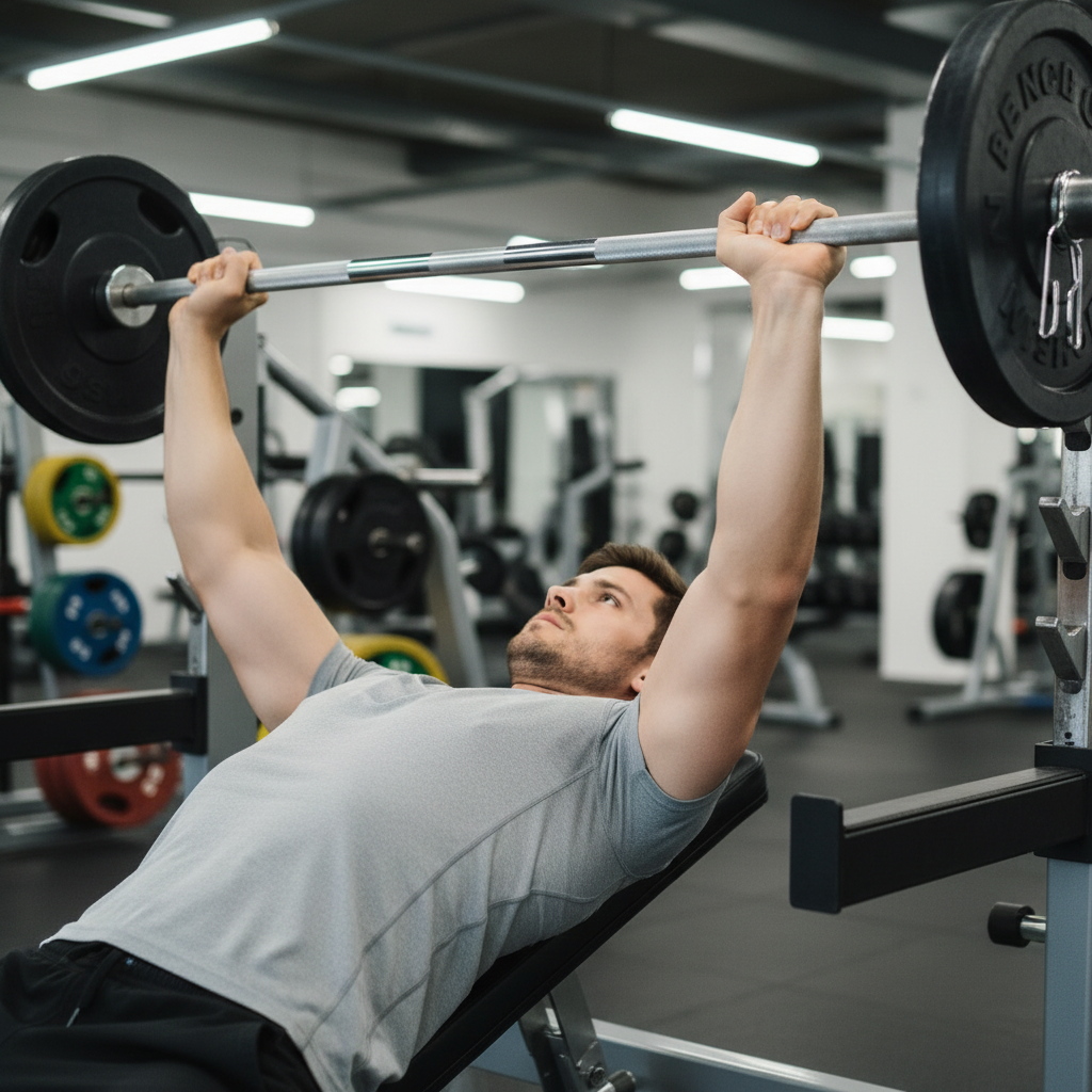
Shoulder pain is one of the most common musculoskeletal complaints, and the causes are more varied than most people expect. This guide walks through the anatomy, the conditions behind your symptoms, how a diagnosis is reached, and an evidence-based path back to pain-free movement.
Anatomy
Why the Shoulder Is Different
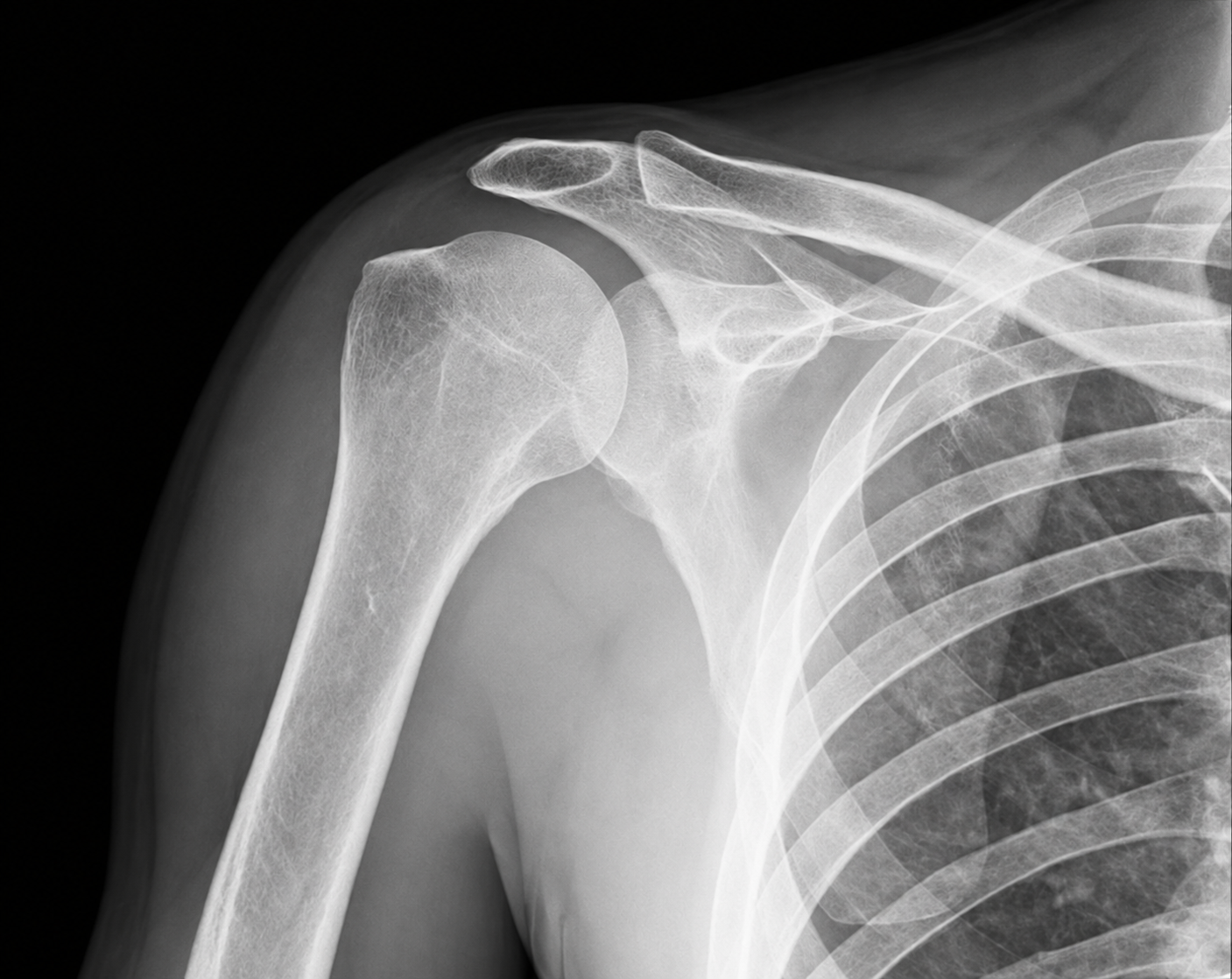
The glenohumeral joint is a shallow ball-and-socket — a large humeral head resting against a small, flat glenoid. This design trades bony stability for extraordinary mobility, making the shoulder the most mobile joint in the body and the most reliant on coordinated soft tissue for control (Ludewig PM & Reynolds JF, J Orthop Sports Phys Ther, 2009).
Because bony contact contributes so little, stability depends almost entirely on the surrounding structures. When any of them is irritated, weakened, or torn, the whole system loses balance — which is precisely why the shoulder is so injury-prone.
The Four Key Structures
- Rotator Cuff — supraspinatus, infraspinatus, teres minor, and subscapularis, which centre and rotate the humeral head.
- Glenoid Labrum — a rim of cartilage that deepens the socket and improves stability.
- Biceps Long Head Tendon — attaches inside the joint and contributes to anterior stability.
- AC Joint — links the shoulder blade to the collarbone and transmits load from the arm.
Common Conditions
What’s Actually Causing Your Pain
Shoulder pain has many faces. Getting the right label matters — because the wrong rehab for the wrong diagnosis can stall progress for months. Here is what each of the major conditions actually involves.
Rotator Cuff Tear / Tendinopathy
The most common cause of shoulder pain in adults over 40. Ranges from tendon fraying to full-thickness tears, most of which are degenerative. Key symptom: pain with overhead activity and night pain (Minagawa H et al., J Orthop Sci, 2013).
Subacromial Impingement Syndrome
Pain arising as tissues are compressed beneath the acromion during arm elevation. Increasingly understood as tendon-related rather than pure mechanical compression. Key symptom: a painful arc between 60 and 120 degrees (Lewis J, Man Ther, 2011).
Adhesive Capsulitis (Frozen Shoulder)
Progressive fibrosis and contracture of the joint capsule causing global stiffness. Disproportionately affects people with diabetes. Key symptom: progressive stiffness in all planes of movement (Hsu JE et al., J Am Acad Orthop Surg, 2011).
Labral Tears (SLAP / Bankart)
Tears of the cartilage rim, common in overhead athletes from repetitive loading (SLAP) or after dislocation (Bankart). Key symptom: clicking, catching, and a sense of instability (Maffet MW et al., Am J Sports Med, 1995).
AC Joint Sprain / Arthritis
Sprain from a direct fall or degenerative arthritis at the joint between collarbone and shoulder blade. Key symptom: pain directly over the AC joint, worse with cross-body movements (Fraser-Moodie JA et al., J Bone Joint Surg Br, 2008).
Biceps Long Head Tendinopathy
Irritation of the proximal biceps tendon inside the joint, frequently alongside rotator cuff pathology. Key symptom: anterior shoulder pain with positive Speed’s and Yergason’s tests (Nho SJ et al., Am J Sports Med, 2010).
Glenohumeral Instability
Traumatic instability follows a dislocation; atraumatic instability arises from generalised hypermobility. Key symptom: a sense of the shoulder slipping, most common in young athletes (Owens BD et al., Am J Sports Med, 2007).
What to Expect
Realistic Recovery Timelines
Healing is not linear. Most people plateau, have flare-ups, and mistake slower weeks for failure. These are evidence-informed ranges — individual variation is significant.
| Condition | Conservative Rehab | Post-Surgical (if applicable) |
|---|---|---|
| Subacromial Impingement | 6–12 weeks | N/A |
| Partial Rotator Cuff Tear | 8–16 weeks | N/A |
| Full-Thickness Rotator Cuff Tear | 12–24 weeks (conservative) | 6–12 months |
| Frozen Shoulder (Adhesive Capsulitis) | 12–24 months | 3–6 months (post-manipulation) |
| Labral Tear (SLAP) | 12–16 weeks (conservative) | 4–6 months |
| AC Joint Separation (Grade I–II) | 4–8 weeks | 3–4 months (Grade III+) |
Moosmayer S et al. Prevalence and characteristics of asymptomatic tears of the rotator cuff. JBJS. 2009. | Milgrom C et al. Rotator cuff tears in cadavers. Clin Orthop Relat Res. 1995.
Surgical Considerations
When Is Surgery Actually Needed?
The evidence is clear: most shoulder conditions resolve with well-directed conservative care. Surgery is indicated in specific circumstances — and the timing matters.
Surgery Is Likely Indicated When:
- Full-thickness rotator cuff tear in active individuals under 65 with acute onset
- Complete tendon rupture (subscapularis, biceps)
- Bankart lesion after recurrent instability episodes (especially under age 25)
- SLAP tear unresponsive to 3–6 months of rehab in overhead athletes
- Frozen shoulder not responding to 6+ months of physio and injections
- Subacromial pain syndrome after 6+ months of structured conservative care (evidence is mixed — shared decision making applies)
Monk AP et al. Evidence for conservative management of subacromial impingement. BJSM. 2017.
Surgery Is Likely NOT Needed When:
- Partial rotator cuff tear (most respond to 12–16 weeks of targeted rehab)
- Subacromial impingement without structural failure
- Frozen shoulder in Stage 1–2 (inflammatory phase)
- AC joint sprain Grade I or II
- Biceps tendinopathy without tendon rupture
- Any shoulder condition that hasn’t completed a full, structured rehab trial
Beard DJ et al. Arthroscopic subacromial decompression for subacromial pain. Lancet. 2018;391(10118):329–338.
Safety First
Red Flags — Seek Urgent Assessment
These symptoms require prompt medical evaluation and should not be managed with self-directed rehab alone:
- Sudden, severe shoulder pain with inability to raise the arm — may indicate acute rotator cuff rupture
- Shoulder pain following trauma with visible deformity — suspect fracture or dislocation
- Pain radiating down the arm with numbness/tingling — cervical radiculopathy or thoracic outlet
- Night pain that is severe, constant, and unresponsive to position change — rule out malignancy
- Systemic symptoms (fever, weight loss, fatigue) with shoulder pain — red flag for systemic pathology
- Rapidly progressive weakness — possible neurological emergency
Ready to Build a Stronger Shoulder?
Evidence-based rehab works — but it works best when it’s matched to your specific diagnosis, stage, and goals. Start with a thorough assessment.
Condition-Specific Guidance
Your Diagnosis Changes Your Protocol
Generic shoulder exercises help — but the most effective rehab is matched to your specific diagnosis. Here’s how each condition modifies the standard protocol.
Frozen Shoulder (Adhesive Capsulitis)
Timeline: 12–24 months without treatment | 6–12 months with physio
Aggressive stretching early worsens inflammation. Phase 1 focuses on pain-free pendulums and heat before movement. Mobilisation is progressed slowly — the capsule must be stretched, not torn.
Key modifications:
- Avoid aggressive end-range stretching in Stage 1
- Heat application 10 min before ROM work
- Hydrodistension may accelerate Phase 2 entry
- Corticosteroid injection most effective within first 6 weeks
Challoumas D et al. Frozen shoulder: an evidence-based approach. EFORT Open Rev. 2022;7(8):553–567.
Rotator Cuff Tear
Timeline: Partial tears 8–12 weeks | Full-thickness (non-surgical) 3–6 months | Post-surgical 6–12 months
Partial tears often respond well to conservative rehab. Full-thickness tears in active individuals under 65 may require surgical repair — outcome is strongly linked to time from injury to surgery.
Key modifications:
- Load progression is slower — tendons repair at ~70% strength at 12 weeks
- Avoid painful arc (60–120° elevation) until Phase 3
- ER strengthening is the single most important exercise target
- Post-surgical: strict non-weight-bearing sling for 4–6 weeks
Pedowitz RA et al. AAOS clinical practice guideline on optimizing the management of rotator cuff problems. JAAOS. 2011.
Subacromial Impingement
Timeline: 6–12 weeks with physio | 80% resolve without surgery
Impingement is largely a load management issue. Scapular control and rotator cuff strengthening reduce the mechanical compression that drives symptoms. Manual therapy accelerates early progress.
Key modifications:
- Scapular upward rotation is the primary biomechanical target
- Avoid overhead loading until pain-free to 120°
- Posterior capsule stretching critical (sleeper stretch)
- Subacromial corticosteroid injection effective short-term for pain control
Steuri R et al. Effectiveness of conservative interventions including exercise, manual therapy and medical management in adults with shoulder impingement. BJSM. 2017.
Rehabilitation
Step-by-Step Rehab Protocol
Shoulder rehab is not one-size-fits-all — but most evidence-based programs progress through four phases, adjusting load and complexity as pain settles and function returns. (Kuhn JE. Exercise in the treatment of rotator cuff impingement. J Shoulder Elbow Surg. 2009;18(1):138–160.)
Phase 1 — Pain Control & Protection
Weeks 1–3 | Goal: Reduce irritability, restore resting comfort
The priority here is calming the tissue — not pushing through pain. Activity modification, relative rest, and gentle movement within a pain-free range are the foundation.
Key Exercises:
- Pendulum swings — 2×20 reps, gravity-assisted, pain-free
- Supine passive flexion — 3×10, using the uninvolved arm to guide
- Scapular retraction — 3×15, gentle, seated
- Cervical mobility — gentle rotation and side-bend, 2×10 each direction
Littlewood C et al. Rehabilitation for rotator cuff tendinopathy. Cochrane Database Syst Rev. 2016.
Phase 2 — Restore Range of Motion
Weeks 3–6 | Goal: Full passive and active ROM in all planes
As pain settles, progressive ROM work begins. Emphasis is on restoring elevation, external rotation, and internal rotation without compensatory patterns.
Key Exercises:
- Pulley-assisted overhead flexion — 3×15, pain-free arc
- Sleeper stretch — 3×30 sec holds, posterior capsule
- Cross-body stretch — 3×30 sec, horizontal adduction
- Active-assisted ER with stick — 3×15 reps
- Wall walks — 3×10, forward and lateral
McClure P et al. Shoulder function and 3-dimensional kinematics in people with shoulder impingement. Phys Ther. 2006.
Phase 3 — Rotator Cuff Strengthening
Weeks 6–12 | Goal: Build rotator cuff and scapular strength
This is where most of the functional gains happen. Strengthening targets the rotator cuff in isolation first, then integrates it into multi-joint patterns. Pain should not exceed 3/10 during exercise.
Key Exercises:
- Side-lying external rotation — 3×15, light dumbbell (1–3 kg)
- Standing ER with band — 3×20, elbow at 90°
- Prone Y-T-W — 3×12 each, scapular control
- Sidelying IR with dumbbell — 3×15
- Scaption (empty can) — 3×15, 30° forward of frontal plane
- Serratus anterior wall push-up plus — 3×15
Reinold MM et al. Electromyographic analysis of the rotator cuff and deltoid musculature. Am J Sports Med. 2004.
Phase 4 — Return to Function & Sport
Weeks 12+ | Goal: Full strength, power, and sport-specific confidence
The final phase integrates overhead loading, plyometrics, and sport-specific patterns. Return-to-sport criteria: pain-free full ROM, strength ≥90% of contralateral side, and confident overhead loading.
Key Exercises:
- Overhead press progression — 3×10, increasing load weekly
- Cable diagonal patterns (PNF D1/D2) — 3×12 each
- Plyometric wall throws — 3×15, medicine ball
- Pull-up progression — 3×8, scapular depression focus
- Sport-specific drills — throwing, swimming, serving as applicable
Wilk KE et al. Shoulder injuries in the overhead athlete. J Orthop Sports Phys Ther. 2009.
Diagnosis
Getting the Right Diagnosis
Diagnosis is a process of clinical reasoning — combining a careful history, a hands-on examination, targeted special tests, and, only when it changes management, imaging. No single test is definitive, so findings are interpreted together.
Clinical History
A structured history points toward the diagnosis before hands ever touch the shoulder. Key questions cover:
- Onset and mechanism of injury
- Night pain and pain at rest
- Sense of instability or slipping
- Occupation and overhead demands
- Sport and repetitive loading
Special Tests
- Neer’s Impingement Sign — Sens 72%, Spec 60% (subacromial impingement)
- Hawkins-Kennedy Test — Sens 79%, Spec 59% (impingement / rotator cuff)
- Empty Can / Full Can Test — Sens 69%, Spec 66% (supraspinatus tear)
- External Rotation Lag Sign — Sens 56%, Spec 98% (full-thickness rotator cuff tear)
- O’Brien’s Active Compression Test — Sens 63%, Spec 73% (SLAP lesion)
- Speed’s Test — Sens 54%, Spec 81% (biceps tendinopathy)
- Apprehension / Relocation Test — Sens 72%, Spec 96% (glenohumeral instability)
Hegedus EJ et al. Physical examination tests of the shoulder: a systematic review with meta-analysis. BJSM. 2012;46(14):964–978.
Imaging: When Is It Useful? Imaging is rarely the first step. Plain X-rays help rule out fracture, calcification, and AC joint arthritis. Diagnostic ultrasound is excellent for dynamic assessment of the rotator cuff and bursa. MRI is indicated when a full-thickness rotator cuff tear, labral pathology, or surgical planning is suspected. Findings should always be interpreted in clinical context — asymptomatic rotator cuff tears are present in 34% of adults over 60 (Tempelhof S et al., JSES 1999).
Cited Sources
1. Kuhn JE. Exercise in the treatment of rotator cuff impingement. J Shoulder Elbow Surg. 2009;18(1):138–160.
2. Hegedus EJ et al. Physical examination tests of the shoulder: a systematic review with meta-analysis. BJSM. 2012;46(14):964–978.
3. Tempelhof S et al. Age-related prevalence of rotator cuff tears in asymptomatic shoulders. J Shoulder Elbow Surg. 1999;8(4):296–299.
4. Littlewood C et al. Rehabilitation for rotator cuff tendinopathy. Cochrane Database Syst Rev. 2016.
5. Challoumas D et al. Frozen shoulder: an evidence-based approach to diagnosis and treatment. EFORT Open Rev. 2022;7(8):553–567.
6. Steuri R et al. Effectiveness of conservative interventions including exercise, manual therapy and medical management in adults with shoulder impingement. BJSM. 2017;51(18):1340–1347.
7. Reinold MM et al. Electromyographic analysis of the rotator cuff and deltoid musculature during common shoulder external rotation exercises. J Orthop Sports Phys Ther. 2004;34(7):385–394.
8. Wilk KE et al. Shoulder injuries in the overhead athlete. J Orthop Sports Phys Ther. 2009;39(2):38–54.
9. Beard DJ et al. Arthroscopic subacromial decompression for subacromial shoulder pain (CSAW). Lancet. 2018;391(10118):329–338.
10. Monk AP et al. Evidence for conservative management of subacromial impingement. BJSM. 2017;51(18):1283–1291.
 Dr. Austin Fontenot