Spinal Arthritis
Evidence-based guides to osteoarthritis, stenosis, flare management, and building a sustainable activity plan.
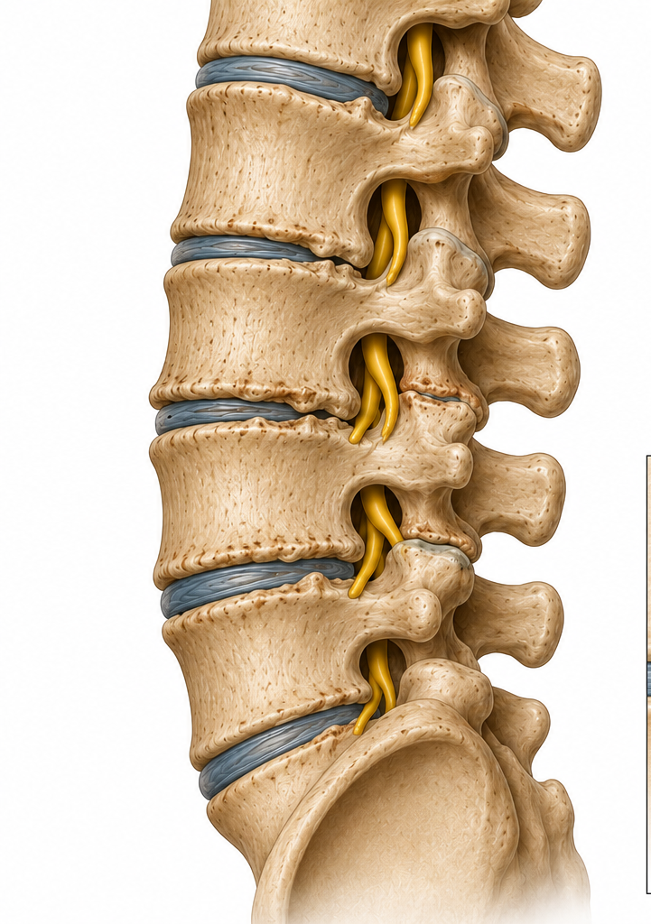
Understanding Spinal Arthritis
Spinal arthritis is an umbrella term for degenerative and inflammatory conditions affecting the joints, cartilage, and surrounding structures of the spine. Two forms dominate the clinical picture: osteoarthritis (OA) — the mechanical wear-and-tear type — and spinal stenosis, often its downstream consequence. Understanding the biology behind each condition is the first step toward managing them intelligently.
What Is Happening
In OA, articular cartilage between facet joints gradually breaks down. Bone-on-bone contact triggers inflammation, osteophyte (bone spur) formation, and joint capsule thickening. The disc may also narrow, compressing adjacent structures.
Who Is Affected
Spinal OA affects an estimated 30–50% of adults over 50, rising sharply with age. Risk factors include prior injury, repetitive loading occupations, obesity, and genetic predisposition. It is not an inevitable consequence of aging — modifiable factors matter enormously.
Why It Matters
Left unmanaged, spinal arthritis can progress to spinal stenosis — narrowing of the spinal canal — causing nerve compression, radiating pain, and neurogenic claudication. Early, evidence-based intervention changes the trajectory.
Two Conditions, One Framework
Osteoarthritis and stenosis share overlapping biology but present differently. Knowing which you are dealing with — or if both are present — shapes your treatment and activity plan.
Most Common Type
Osteoarthritis (OA)
OA of the spine involves progressive breakdown of the facet joint cartilage and intervertebral discs. Hallmarks include morning stiffness lasting under 30 minutes, pain that worsens with sustained postures, crepitus, and bony enlargement on imaging. Pain is typically axial (local to the spine) though referred pain to the buttocks or thighs is common. Activity-pacing, load management, and targeted exercise are the cornerstones of OA management. Evidence consistently shows that movement — done right — slows progression.
Often OA’s Downstream Effect
Spinal Stenosis
Stenosis refers to narrowing of the spinal canal or neural foramina, often resulting from OA-related osteophytes, ligamentum flavum thickening, or disc bulging. Classic presentation is neurogenic claudication: leg heaviness, cramping, or pain brought on by walking or standing that is relieved by sitting or flexion. Lumbar stenosis affects roughly 11% of adults over 60. Management follows similar principles to OA — exercise, activity modification, and targeted therapies — with additional attention to neural tension and posture.
Managing Flares: The Evidence-Based Approach
Flares are temporary intensifications of arthritis symptoms — increased pain, swelling, stiffness, and fatigue. They are not signs of permanent damage. The goal during a flare is to reduce load while preserving movement, not to stop moving entirely.
During a Flare
Reduce but don’t eliminate activity. Gentle range-of-motion exercises, walking, and aquatic therapy are preferred. Apply ice or heat (patient preference — both show similar efficacy). Avoid prolonged bed rest: immobilization accelerates joint stiffness and muscle loss. (Fransen et al., Cochrane Review, 2015)
Activity Pacing
Pace rather than push. Break tasks into shorter intervals with rest periods. Use a ‘boom-bust’ awareness approach: identify your personal threshold and stay just below it consistently. Energy conservation during flares preserves capacity for recovery. (Gill & Brown, 2009, Pain Management Nursing)
Post-Flare Return
Gradually increase activity within 48–72 hours of symptom improvement. Begin with aquatic or gravity-reduced exercise before returning to land-based loading. Expect 1–2 weeks to return to baseline. Track flare triggers (weather, activity type, stress) to inform future pacing.
Conventional Treatment Options
Conservative care remains first-line for spinal arthritis. The evidence strongly favors a multimodal approach over any single intervention.
NSAIDs
Non-steroidal anti-inflammatory drugs (ibuprofen, naproxen, celecoxib) are first-line pharmacological treatment for OA pain. Short-term use is generally well-tolerated; long-term use requires GI and cardiovascular monitoring. (Hochberg et al., ACR Guidelines, 2012)
Corticosteroid Injections
Intra-articular or epidural corticosteroid injections provide short-term pain relief (4–8 weeks) and can enable participation in rehabilitation. Repeated injections (>3–4/year) carry cartilage degradation risk and are not recommended as a long-term strategy. (Manchikanti et al., Pain Physician, 2013)
Physical Therapy
Structured PT programs combining manual therapy, therapeutic exercise, and patient education demonstrate superior long-term outcomes compared to passive modalities alone. Emphasis on progressive loading and functional movement patterns. (Deyle et al., NEJM Evidence, 2020)
Hyaluronic Acid Injections
Viscosupplementation (e.g., Synvisc, Euflexxa) may offer modest pain relief in knee OA; evidence for spinal application is limited. Best considered when NSAIDs are contraindicated. (Bannuru et al., Osteoarthritis & Cartilage, 2019)
Topical Analgesics
Topical diclofenac and capsaicin provide localized pain relief with minimal systemic effects — a good option for those who cannot tolerate oral NSAIDs. Evidence strongest for peripheral joint OA but applicable to paraspinal soft tissue pain. (Derry et al., Cochrane, 2016)
Surgical Options
Surgery (laminectomy, spinal fusion) is reserved for cases with significant neurological compromise or failed conservative care over 6–12 months. Surgical outcomes for pain relief alone are modest; outcomes for neurological symptoms are generally better. (Weinstein et al., SPORT Trial, JAMA, 2008)
Therapeutic Exercise
Exercise is the single most evidence-supported intervention for spinal arthritis. It reduces pain, improves function, slows structural progression, and supports mental health. The key is matching exercise type and intensity to your current stage — not avoiding movement out of fear.
Aquatic Exercise
Water-based exercise reduces joint load by up to 90% in waist-deep water, allowing pain-free movement when land-based loading is too painful. Cochrane reviews confirm aquatic therapy significantly reduces pain and improves function in OA. Ideal for flare periods and initial rehabilitation. (Bartels et al., Cochrane, 2016)
Resistance Training
Strengthening the muscles surrounding the spine — erector spinae, multifidus, gluteals, hip flexors — reduces compressive load on arthritic facets. Progressive resistance training 2–3×/week is strongly recommended. Begin with bands before progressing to free weights. (Fransen et al., Cochrane, 2015)
Core Stabilization
Motor control exercises — dead bugs, bird dogs, pallof press — retrain deep stabilizers (transversus abdominis, multifidus) and significantly reduce pain in lumbar OA. A dysfunctional stabilizer system accelerates arthritic joint loading. (Saragiotto et al., Cochrane, 2016)
Walking & Aerobic
Moderate aerobic activity (150 min/week, WHO) reduces systemic inflammation, supports cartilage nutrition via synovial fluid circulation, and improves body composition. Walking, cycling, and elliptical training are all appropriate low-impact options. (Roddy et al., Ann Rheum Dis, 2005)
Tai Chi & Mind-Body
Tai chi combines slow controlled movement with breathing and balance training. RCTs demonstrate significant reductions in pain, stiffness, and fear of movement vs. control groups. Particularly effective in older adults with stenosis. (Wang et al., Arthritis Care Res, 2009)
Stretching & Mobility
Hip flexor, hamstring, and thoracic mobility work reduces compensatory lumbar loading. Hold stretches 30–60 seconds × 3 reps daily. Neural mobilization (nerve flossing) addresses radicular symptoms in stenosis patients.
Laser Therapy for Spinal Arthritis
Photobiomodulation (PBM) — delivered via Class IV or Class IIIb low-level laser — is an evidence-backed modality for arthritis pain. It works by stimulating mitochondrial activity, reducing inflammatory cytokines, and accelerating tissue repair at the cellular level.
Mechanism of Action
Near-infrared wavelengths (810–1064 nm) penetrate deep tissue, activating cytochrome c oxidase in mitochondria. This triggers increased ATP production, reduced oxidative stress, and modulation of pro-inflammatory cytokines (TNF-α, IL-1β, IL-6) — all relevant to the arthritic joint environment. (Hamblin, SPIE, 2017)
Clinical Evidence
A systematic review and meta-analysis of 16 RCTs found LLLT significantly reduces pain and morning stiffness in musculoskeletal OA. The World Association for Laser Therapy (WALT) has published dosing guidelines specifically for OA. (Bjordal et al., BMC Musculoskelet Disord, 2003)
Treatment Protocol
Typical protocols involve 6–12 sessions over 3–6 weeks, targeting facet joints and periarticular tissue. Class IV lasers (3–25W) allow deeper penetration for lumbar targets. Best outcomes occur when combined with therapeutic exercise and manual therapy.
Chiropractic Care for Spinal Arthritis
Chiropractic care offers several evidence-supported interventions for spinal arthritis — primarily for pain reduction, mobility restoration, and functional improvement. Importantly, technique selection must account for the degree of degeneration: high-velocity manipulation is modified or avoided in advanced stenosis or severe osteoporosis.
Spinal Mobilization
Low-velocity, controlled joint mobilization techniques (grades I–III) are appropriate for arthritic spines. They improve joint fluid distribution, reduce capsular adhesions, and modulate pain via neurological gating mechanisms. (Bronfort et al., Best Practice & Research, 2008)
Soft Tissue Therapy
Myofascial release, trigger point therapy, and instrument-assisted soft tissue mobilization (IASTM) address the compensatory muscle guarding that accompanies chronic arthritis. Reduces pain and improves range of motion without loading the arthritic joint directly.
Flexion-Distraction
A gentle, non-thrust technique particularly useful in lumbar stenosis. Flexion-distraction increases disc height, widens the spinal canal, and reduces intradiscal pressure. Studies show significant reduction in leg pain and neurogenic claudication. (Cox & Shreiner, JMPT, 1992)
Therapeutic Ultrasound & E-Stim
Adjunct modalities including therapeutic ultrasound and electrical stimulation (TENS/interferential) are commonly used to reduce periarticular inflammation and improve local circulation. Best used in combination with active rehabilitation rather than as standalone treatments.
Building a Sustainable Activity Plan
A sustainable activity plan for spinal arthritis is not about doing more — it is about doing the right things, in the right sequence, with the right recovery built in. The four-step framework below provides a structured approach grounded in progressive loading principles and clinical evidence.
Step 1: Establish Your Baseline
Before adding load, identify your pain-free range of motion, your walking tolerance, and your daily step count. This is your floor, not your ceiling. Track for 1–2 weeks before progressing.
Step 2: Progressive Loading
Increase volume or intensity by no more than 10% per week. Prioritize frequency over intensity early on. Progress from aquatic → gravity-reduced → full weight-bearing → resistance training as tolerance improves.
Step 3: Recovery Integration
Sleep, nutrition, and stress management are part of the plan. Poor sleep elevates inflammatory markers. Chronic stress accelerates cartilage breakdown. Build in 1–2 full rest days per week.
Step 4: Monitor & Adjust (The 24-Hour Rule)
If pain is significantly worse the morning after a session, the load was too high. Mild soreness resolving within 24 hours is acceptable. Adjust every 4–6 weeks based on step count, pain scores, and sit-to-stand time.
When to Seek Medical Attention
Spinal arthritis is manageable for most people. However, certain symptoms indicate nerve compromise or systemic disease requiring urgent evaluation.
Schedule a Visit
- Pain persisting beyond 6 weeks despite conservative care
- Increasing stiffness limiting daily activities
- New onset leg pain or cramping with walking
- Unexplained fatigue or weight loss alongside joint pain
Seek Prompt Evaluation
- Progressive leg weakness or numbness
- Loss of balance or frequent falls
- Radiating pain below the knee that is new or worsening
- Significant functional decline
⚠️ Seek Emergency Care
- Loss of bowel or bladder control
- Saddle anesthesia (numbness in groin/inner thighs)
- Rapid onset bilateral leg weakness
These may indicate cauda equina syndrome — a surgical emergency.
Nutrition for Spinal Arthritis
What you eat directly influences systemic inflammation, joint tissue health, and body weight — three of the most modifiable drivers of arthritis progression. An anti-inflammatory diet is not a cure, but the evidence supporting its role in symptom management is substantial.
Mediterranean Diet
The Mediterranean diet — rich in olive oil, fatty fish, vegetables, legumes, and whole grains — is the most evidence-backed dietary pattern for reducing inflammatory markers (CRP, IL-6) in OA. Adherence is associated with lower pain scores and slower radiographic progression. (Veronese et al., Nutrients, 2022)
Omega-3 Fatty Acids
EPA and DHA (found in fatty fish and quality fish oil supplements) inhibit prostaglandin synthesis and reduce synovial inflammation. Meta-analyses support supplementation at 2–4g/day for measurable reductions in joint pain and stiffness. (Goldberg & Katz, Pain, 2007)
Weight Management
Each kilogram of body weight reduction removes approximately 4 kg of compressive force from the lumbar spine. A 10% body weight reduction produces clinically significant improvements in pain and function in spinal OA. Caloric deficit combined with resistance training is the most effective strategy. (Christensen et al., Ann Rheum Dis, 2007)
Key Nutrients
Vitamin D (deficiency worsens OA pain; target 40–60 ng/mL), magnesium (involved in 300+ enzymatic reactions including muscle relaxation), and collagen peptides (10–15g/day with vitamin C may support cartilage matrix synthesis) are among the most studied. (McAlindon et al., JAMA, 2019; Shaw et al., Am J Clin Nutr, 2017)
Sources & References
This page draws on peer-reviewed research, clinical practice guidelines, and systematic reviews. Key sources are listed below.
Bartels EM, et al. Aquatic exercise for the treatment of knee and hip osteoarthritis. Cochrane Database Syst Rev. 2016.
Bjordal JM, et al. A systematic review of low level laser therapy with location-specific doses for pain from chronic joint disorders. BMC Musculoskelet Disord. 2003.
Bronfort G, et al. Effectiveness of manual therapies: the UK evidence report. Chiropr Osteopat. 2010.
Christensen R, et al. Effect of weight reduction in obese patients diagnosed with knee osteoarthritis. Ann Rheum Dis. 2007.
Cox JM, Shreiner S. Chiropractic manipulation in low back pain and sciatica. JMPT. 1992.
Deyle GD, et al. Physical therapy versus glucocorticoid injection for osteoarthritis of the knee. NEJM Evidence. 2020.
Derry S, et al. Topical NSAIDs for chronic musculoskeletal pain in adults. Cochrane Database Syst Rev. 2016.
Fransen M, et al. Exercise for osteoarthritis of the knee. Cochrane Database Syst Rev. 2015.
Gill JR, Brown CA. A structured review of the evidence for pacing as a chronic pain intervention. Pain Management Nursing. 2009.
Goldberg RJ, Katz J. A meta-analysis of the analgesic effects of omega-3 polyunsaturated fatty acid supplementation for inflammatory joint pain. Pain. 2007.
Hamblin MR. Mechanisms and applications of the anti-inflammatory effects of photobiomodulation. AIMS Biophys. 2017.
Hochberg MC, et al. American College of Rheumatology 2012 recommendations for use of nonpharmacologic and pharmacologic therapies in osteoarthritis. Arthritis Care Res. 2012.
Manchikanti L, et al. A systematic review of effectiveness of therapeutic facet joint interventions in managing chronic spinal pain. Pain Physician. 2013.
Roddy E, et al. Aerobic walking or strengthening exercise for osteoarthritis of the knee? Ann Rheum Dis. 2005.
Saragiotto BT, et al. Motor control exercise for chronic non-specific low-back pain. Cochrane Database Syst Rev. 2016.
Veronese N, et al. Mediterranean diet and osteoarthritis: a systematic review. Nutrients. 2022.
Wang C, et al. Tai Chi is effective in treating knee osteoarthritis: a randomized controlled trial. Arthritis Care Res. 2009.
Weinstein JN, et al. Surgical versus nonsurgical therapy for lumbar spinal stenosis (SPORT). JAMA. 2008.
Arthritis Doesn’t Have to Define Your Movement
The evidence is clear: the right combination of movement, treatment, and nutrition can meaningfully reduce pain, slow progression, and restore quality of life. Explore more condition-specific guides below.
 Dr. Austin Fontenot