Hip Health
Evidence-based guidance on hip pain, mobility, strength, and common conditions affecting everyday movement.
Understanding Hip Function and Pain
The hip is a powerful ball-and-socket joint that lets you walk, squat, and rotate. When muscles, tendons, or cartilage become irritated, pain may appear in the groin, side of the hip, buttock, or down the leg.
Why the Hip Is Built the Way It Is
The hip is a ball-and-socket joint formed by the femoral head and the acetabulum of the pelvis. This architecture allows an extraordinary range of motion — flexion, extension, abduction, adduction, and rotation — while bearing loads several times body weight with each step.
Key Structures
- Labrum: fibrocartilaginous ring deepening the socket, adding ~22% contact area
- Iliofemoral ligament: strongest ligament in the human body, resists hip extension
- Hip joint capsule: surrounds and stabilizes the joint
- Iliopsoas (hip flexors): primary hip flexor, critical for gait
- Gluteus maximus: primary hip extensor, largest muscle in the body
- Gluteus medius & minimus: hip abductors, pelvis stabilizers during single-leg stance
- Deep external rotators (piriformis, obturators, gemelli, quadratus femoris): fine-tune femoral head position
- TFL / IT band complex: lateral hip and knee stabilizer
Why Hip Strength Matters Beyond the Hip
Weak glutes and hip stabilizers don’t just cause hip pain — they alter mechanics at the knee and lower back. Trendelenburg gait (pelvis drop during single-leg stance) from gluteus medius weakness is strongly associated with patellofemoral pain, IT band syndrome, and lumbar overload.
Neumann DA. Kinesiology of the Musculoskeletal System. 3rd ed. Elsevier; 2017.
Getting the Right Diagnosis
Accurate diagnosis requires combining your history, physical examination findings, and selective imaging. Pain location is the starting point — but referred pain patterns mean the source is often not where you feel it.
Clinical History — What Clinicians Ask
- Where exactly is the pain? (groin = intra-articular; lateral = GTPS/tendon; buttock = SI joint, piriformis, or lumbar)
- Sharp catch vs. dull ache vs. burning?
- What makes it worse? (sitting → FAI/labrum; lying on it → GTPS; walking → OA)
- Any clicking, locking, or giving way?
- Morning stiffness lasting >1 hour? (consider inflammatory arthritis)
- Recent increase in training load or mileage? (stress fracture risk)
- Any history of childhood hip problems? (Perthes, SCFE — increases later OA risk)
Key Physical Examination Tests
| Test | What It Detects | Sensitivity / Specificity |
|---|---|---|
| FABER (Patrick’s) | Hip / SI joint pathology | Sens 60%, Spec 18% (hip); improves with cluster |
| FADIR | FAI / labral pathology | Sens 78%, Spec 10% — high sensitivity, low specificity |
| Log Roll Test | Intra-articular hip pathology | Sens 30%, Spec 90% — high specificity |
| Trendelenburg Sign | Gluteus medius weakness | Sens 55–73%, Spec 77–85% |
| Ober’s Test | TFL / IT band tightness | Clinical utility debated; use with context |
| Resisted Hip Flexion | Iliopsoas tendon pathology | Positive if anterior hip pain reproduced |
| Thigh Thrust | SI joint provocation | Part of validated SI joint cluster |
No single test rules in or out hip pathology. Sensitivity and specificity data from Reiman MP et al. Physical examination tests for hip pathology. Br J Sports Med. 2013.
Imaging — What It Shows and What It Doesn’t
X-Ray (First Line)
AP pelvis + lateral hip view identifies joint space narrowing (OA), cam/pincer morphology (FAI), fractures, and dysplasia. X-ray findings frequently don’t correlate with symptoms — up to 45% of asymptomatic adults show cam morphology.
Frank JM et al. Prevalence of FAI findings on skeletal survey radiographs. Am J Sports Med. 2015.
MRI / MR Arthrogram
Standard MRI assesses soft tissue (labrum, cartilage, tendons, stress fractures). MR arthrogram (gadolinium injected into the joint) significantly improves labral tear detection sensitivity (90–95% vs. 30% for standard MRI). Use when surgical planning is being considered.
Chopra A et al. MR arthrography of the hip. RadioGraphics. 2018.
Ultrasound
Excellent for dynamic assessment of tendon pathology, bursae, and snapping hip. Allows real-time assessment of the iliopsoas or IT band during movement. Also used to guide injections into the trochanteric bursa or hip joint.
When NOT to Image First
Most hip pain doesn’t need imaging at the first visit. A 4–6 week trial of conservative management (load modification, targeted exercise) is appropriate for suspected tendinopathy, mild OA, or muscle-related pain. Image immediately if: trauma with suspected fracture, red flags (see below), or failure to improve with appropriate care.
Step-by-Step Hip Rehabilitation Protocol
Hip rehab isn’t one-size-fits-all — a labral tear has different loading priorities than GTPS or OA. The phases below reflect evidence-based principles applicable across most hip conditions, with condition-specific notes where the approach diverges. Progress is guided by symptoms, not just time.
Based on: Lewis CL et al. Effect of hip angle on gluteus maximus recruitment. J Electromyogr Kinesiol. 2009. | Distefano LJ et al. Gluteal muscle activation during common therapeutic exercises. J Orthop Sports Phys Ther. 2009.
Phase 1 — Pain Control & Load Management (Weeks 1–3)
The goal is not rest — it’s relative rest. Eliminate provocative loads (for GTPS: no crossing legs, no lying on affected side; for FAI: avoid deep hip flexion past 90°; for OA: reduce high-impact loading) while maintaining circulation and gentle neuromuscular activation.
Key Exercises
- Supine heel slides — 3×15 reps, pain-free range only
- Sidelying hip abduction (non-compressive position) — 3×15 reps
- Clamshells with resistance band — 3×20 reps
- Seated hip flexion march — 3×15 each side
- Prone hip extension — 3×15 reps
- Ankle pumps + gentle hip circles — 2×20 reps
What to Avoid in Phase 1
- Crossing legs (GTPS provocation)
- Deep squats below 90° hip flexion (FAI/labral irritation)
- High-impact activity (running, jumping)
- Aggressive stretching into pain
Outcome Goal
Pain ≤3/10 with ADLs. Able to perform all Phase 1 exercises without symptom reproduction.
Phase 2 — Restore Range of Motion & Neuromuscular Control (Weeks 3–8)
Begin progressive hip mobility work and introduce closed-chain loading. Emphasis shifts to gluteus medius and deep rotator retraining — the muscles most consistently weak in hip pain populations.
Key Exercises
- Standing hip abduction with band — 3×15 each side
- Single-leg stance (eyes open → eyes closed) — 3×30 sec each side
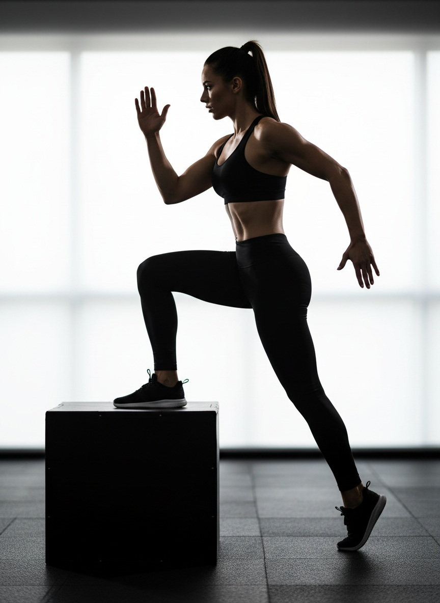
- Step-ups onto 4–8 inch box — 3×12 each side
- Hip flexor stretch (90/90 position, pain-free) — 3×45 sec each side
- Bridge with band above knees — 3×15 reps
- Side-lying hip external rotation — 3×15 reps
- Lateral band walks — 3×15 steps each direction
Progress Criteria
Single-leg stance ≥30 seconds without trunk sway. Pain ≤2/10 during all exercises. Symmetrical hip ROM within 10° of uninvolved side.
Distefano LJ et al. Gluteal muscle activation during therapeutic exercises. J Orthop Sports Phys Ther. 2009.
Phase 3 — Progressive Strength & Functional Loading (Weeks 6–14)
Load the hip through full range with progressive resistance. The evidence strongly favours heavy slow resistance training for tendinopathy and progressive loading for OA. Avoid provocation — some pain (≤4/10 that settles within 24 hours) is acceptable during loading phase.
Key Exercises
- Goblet squat — 4×8–10 reps @ RPE 7/10
- Romanian deadlift (RDL) — 4×8–10 reps, focus on hip hinge
- Hip thrust / barbell glute bridge — 4×10–12 reps
- Lateral step-down — 3×12 each side (eccentric control focus)
- Copenhagen adduction — 3×8–10 each side
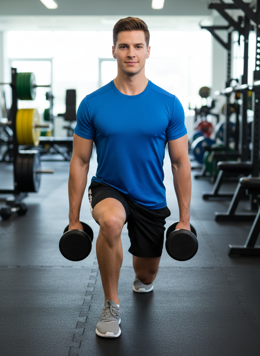
- Bulgarian split squat — 3×10 each side
- Suitcase carry — 3×20m each side (unilateral loading)
Condition-Specific Notes
- GTPS: avoid compressive positions (deep squat, hip adduction past neutral) until symptom-free; prioritize hip abductor loading
- FAI/Labrum: avoid deep impingement angles; bias hip hinge patterns over deep squat
- Hip OA: heavy slow resistance training 3×/week shown to reduce pain and improve function (Svege I et al. Ann Rheum Dis. 2015)
- Hip flexor tendinopathy: eccentric-concentric continuum; start isometric holds before dynamic loading
Svege I et al. Exercise therapy may postpone total hip replacement surgery. Ann Rheum Dis. 2015. | Mellor R et al. Education plus exercise for GTPS. BMJ. 2018.
Phase 4 — Return to Sport & Full Function (Weeks 12+)
Return to sport is guided by functional benchmarks, not a calendar. Symmetry in strength and power, absence of compensatory movement patterns, and psychological readiness all factor in.
Return-to-Sport Criteria
- Hip abductor strength ≥90% of uninvolved side (dynamometry preferred)
- Single-leg hop test ≥90% limb symmetry index
- Y-Balance Test posterior reach within 4 cm of uninvolved side
- Pain ≤1/10 during sport-specific activity
- No antalgia (pain-avoidant gait) during jogging
Progressive Return Timeline
- Week 12–14: Straight-line jogging, light plyometrics
- Week 14–16: Cutting, acceleration/deceleration, sport-specific drills
- Week 16+: Full training, then return to competition
Reiman MP et al. Return to sport after hip and groin injuries. J Orthop Sports Phys Ther. 2017.
Realistic Recovery Timelines
Recovery is rarely linear. The timelines below reflect typical conservative management outcomes. Surgical timelines, high-symptom burden at baseline, and poor adherence to progressive loading can extend these estimates significantly.
| Condition | Conservative Timeline | Key Milestone | Notes |
|---|---|---|---|
| Hip OA (mild–moderate) | 6–12 weeks to meaningful improvement | Pain ≤3/10 with walking | Exercise is first-line; improvement continues with maintenance loading |
| Hip OA (severe) | Ongoing management; THA if failed conservative | Functional walking tolerance | THA outcomes excellent; average surgery age ~65 |
| Femoroacetabular Impingement | 8–16 weeks | Return to sport criteria met | ~60% avoid surgery with structured rehab (Wall PDH et al. BJSM 2016) |
| Hip Labral Tear | 12–16 weeks conservative; 4–6 months post-op | Return to pivoting/sport | Many labral tears are asymptomatic — imaging alone should not drive surgery |
| Greater Trochanteric Pain Syndrome | 8–16 weeks | No pain with lying on side | Compressive load avoidance critical in early phase |
| Hip Flexor Tendinopathy | 8–12 weeks | Pain-free resisted hip flexion | Isometric loading in early phase; eccentric-concentric progression after |
| Piriformis Syndrome | 6–10 weeks | No radicular symptoms | Rule out lumbar contribution; manual therapy + stretching effective |
| Femoral Neck Stress Fracture | 6–12 weeks non-weight-bearing (tension-side) | Radiographic healing confirmed | Urgent orthopaedic referral required; no conservative-only management for tension-side |
| SI Joint Dysfunction | 6–12 weeks | Negative provocation cluster | Manual therapy + stabilization exercises; prolotherapy/injection if refractory |
Timelines adapted from: Bizzini M et al. Recommendations for return to sport after hip and groin injuries. Br J Sports Med. 2012.
What’s Actually Causing Your Hip Pain
Hip pain is a symptom, not a diagnosis. Location matters: groin pain often points to intra-articular pathology (labrum, cartilage, joint), lateral hip pain to tendons or bursae, and buttock pain to the SI joint, deep external rotators, or referred lumbar spine pain. Here are the most common culprits.
Hip Osteoarthritis (OA)
The most common hip condition in adults over 50. Cartilage breakdown leads to groin pain, stiffness after rest, and reduced internal rotation. X-ray findings often don’t match symptoms — radiographic OA is common without pain. Exercise is first-line treatment.
Quicke JG et al. Exercise therapy for hip and knee OA. Cochrane Database Syst Rev. 2015.
Femoroacetabular Impingement (FAI)
Bony overgrowth on the femoral head (cam), acetabular rim (pincer), or both causes mechanical conflict during hip flexion. Common in young active adults. Symptoms: anterior groin pain with deep hip flexion, prolonged sitting, or pivoting. Diagnosis confirmed with X-ray and MRI.
Griffin DR et al. FAI syndrome: moving from a recognition towards a definition. Br J Sports Med. 2016.
Hip Labral Tear
The labrum deepens the socket and seals joint fluid. Tears — often from FAI, trauma, or repetitive pivoting — cause groin pain, clicking, locking, or a feeling of instability. MRI arthrogram is the gold standard for diagnosis. Many labral tears are asymptomatic and found incidentally.
Domb BG et al. Hip labral tears: correlating MRI and arthroscopic findings. Arthroscopy. 2014.
Greater Trochanteric Pain Syndrome (GTPS)
Previously called trochanteric bursitis, GTPS is now understood as primarily a gluteus medius/minimus tendinopathy. Pain on the outer hip, worse with lying on the affected side, crossing legs, or climbing stairs. More common in women. Compressive loading (crossing legs, hip adduction) is a key driver.
Mellor R et al. Education plus exercise for GTPS. BMJ. 2018.
Hip Flexor Tendinopathy / Iliopsoas Syndrome
Repetitive hip flexion (running, cycling, sit-ups) can irritate the iliopsoas tendon where it crosses the iliopectineal eminence. Symptoms: anterior hip pain that worsens with resisted hip flexion, going up stairs, or sitting to standing. A snapping sensation may also be present (coxa saltans interna).
Dydyk AM, Gupta N. Iliopsoas Muscle Injuries. StatPearls. 2023.
Piriformis Syndrome
The piriformis muscle can irritate the sciatic nerve as it exits the greater sciatic foramen. Symptoms mimic lumbar sciatica: deep buttock pain radiating down the posterior thigh. Key differentiator: pain reproduced by hip rotation, not lumbar flexion. Prevalence remains debated; likely over-diagnosed.
Probst D et al. Piriformis Syndrome: A Narrative Review of the Anatomy, Diagnosis, and Treatment. Am J Phys Med Rehabil. 2019.
Stress Fracture of the Femoral Neck
A red flag in runners, military recruits, and those with relative energy deficiency in sport (RED-S). Pain in the groin that worsens with weight-bearing and doesn’t respond to rest. X-ray may be normal; MRI is required for diagnosis. Tension-side stress fractures require urgent orthopaedic referral.
Behrens SB et al. Femoral neck stress fractures. Am J Orthop. 2013.
Sacroiliac (SI) Joint Dysfunction
The SI joint transfers load between the spine and lower limbs. Dysfunction causes buttock pain that may refer into the posterior thigh. Diagnosis is clinical — no single test is definitive, but clusters of provocation tests (FABER, FADIR, Gaenslen’s, thigh thrust) improve accuracy. Often coexists with lumbar pathology.
Laslett M. Evidence-based diagnosis and treatment of the SI joint. J Man Manip Ther. 2008.
Knowing When to Progress in Rehabilitation
Deciding when to progress your hip rehabilitation should be based on objective changes in symptoms, function, and load tolerance—not just the passage of time. Most hip pain conditions, including labral pathology, FAI, mild-to-moderate osteoarthritis (OA), and tendinopathy, respond well to structured conservative management that is appropriately progressed and periodically reassessed (Grimaldi A et al. J Orthop Sports Phys Ther. 2015). Progression should be deliberate, criteria-based, and individualized to your goals and starting point.
Evidence for Conservative Progression
- Hip OA: Supervised exercise and education improve pain, function, and walking capacity in hip OA, with benefits maintained long-term when patients continue home programs (Svege I et al. Ann Rheum Dis. 2015). Progression is typically guided by tolerable soreness (<3/10 that resolves within 24 hours) and gradual increases in walking distance, sit-to-stand repetitions, and loaded tasks.
- FAI: Structured physiotherapy emphasizing hip and lumbopelvic control, strength, and graded exposure to aggravating tasks leads to clinically meaningful improvements for many with mild-to-moderate FAI (Mansell NS et al. Am J Sports Med. 2018). Key progression markers include improved hip range of motion, better control in single-leg tasks, and reduced pain during previously provocative positions.
- GTPS: Education plus progressive loading of the gluteal tendons outperforms passive approaches alone, with superior pain and function outcomes at 8 and 52 weeks (Mellor R et al. BMJ. 2018). Exercise is typically progressed by increasing load, speed, or complexity when pain remains tolerable during activity and settles to baseline within 24 hours.
- Labral pathology: Many people have labral findings on MRI without symptoms, and clinical outcomes often depend more on strength, control, and load management than the imaging itself (Register B et al. Am J Sports Med. 2012). Rehabilitation progresses as hip strength, balance, and confidence improve, and as you can perform daily and sport-specific tasks with only mild, short-lived discomfort.
Clinical Checkpoints for Progression
- Consistent pain levels ≤3/10 during exercise that return to baseline within 24 hours, without increasing night pain or next-day stiffness
- Improved function in key tasks (walking distance, stair climbing, sit-to-stand, single-leg balance) measured over several weeks
- Ability to perform current exercises with good technique and minimal compensation before adding load, speed, range, or complexity
- Stable or improving symptoms over at least 2–4 weeks at a given training level, rather than day-to-day fluctuation without a clear upward trend
- Gradual return to desired activities (work, sport, or recreation) with manageable, short-lived increases in soreness that do not accumulate over the week
When to Seek Further Clinical Evaluation
- Pain that is worsening over several weeks despite consistent, well-performed rehabilitation and appropriate load management
- Persistent night pain, unexplained weight loss, fever, or a history of significant trauma associated with hip pain
- New or increasing difficulty with weight-bearing, such as limping that does not improve, or loss of strength that is out of proportion to pain
- Loss of hip motion, function, or confidence that does not respond to a 6–8 week trial of progressive, criteria-based exercise
- Uncertainty about whether your current program is appropriate, too easy, or too aggressive, or if you are unsure how to progress safely
- Desire to return to higher-level sport or physically demanding work and wanting a structured, criteria-based plan for advancement and risk reduction
Seeking input from a sports medicine physician, physical therapist, or musculoskeletal specialist can help you clarify a diagnosis, optimize your rehabilitation plan, and establish clear progression criteria, especially if your recovery has plateaued or you are unsure about the next steps.
🚨 Red Flags — Seek Urgent Assessment
Most hip pain is benign and musculoskeletal. These signs, however, require prompt medical evaluation and should not be managed with exercise or wait-and-see:
- Severe hip/groin pain after trauma, fall, or impact — possible fracture or dislocation
- Inability to weight-bear after injury
- Fever + joint pain + warmth/swelling — possible septic arthritis (medical emergency)
- Night pain that wakes you from sleep consistently
- Unexplained weight loss with new hip pain
- History of cancer + new persistent hip/groin pain — rule out metastatic disease
- Pain at rest that does not improve with any position
- Rapidly progressive neurological symptoms (weakness, numbness, loss of bladder/bowel)
- Acute severe groin pain in a child or adolescent — rule out Perthes disease or SCFE
- Pulsatile mass or bruit in groin — vascular referral
When in doubt, see a clinician. Early diagnosis changes outcomes for fractures, septic arthritis, and oncological conditions.
Cited References
- Neumann DA. Kinesiology of the Musculoskeletal System. 3rd ed. Elsevier; 2017.
- Quicke JG et al. Exercise therapy for hip and knee osteoarthritis. Cochrane Database Syst Rev. 2015.
- Griffin DR et al. FAI syndrome: moving from a recognition towards a definition. Br J Sports Med. 2016;50(19):1169–1176.
- Domb BG et al. Hip labral tears: correlating MRI and arthroscopic findings. Arthroscopy. 2014.
- Mellor R et al. Education plus exercise versus corticosteroid injection for GTPS. BMJ. 2018;361:k1662.
- Dydyk AM, Gupta N. Iliopsoas Muscle Injuries. StatPearls [Internet]. 2023.
- Probst D et al. Piriformis Syndrome: A Narrative Review. Am J Phys Med Rehabil. 2019;98(9):793–799.
- Behrens SB et al. Femoral neck stress fractures. Am J Orthop. 2013.
- Laslett M. Evidence-based diagnosis and treatment of the painful sacroiliac joint. J Man Manip Ther. 2008;16(3):142–152.
- Reiman MP et al. Physical examination tests for hip pathology. Br J Sports Med. 2013.
- Frank JM et al. Prevalence of FAI findings on skeletal survey radiographs. Am J Sports Med. 2015.
- Chopra A et al. MR arthrography of the hip. RadioGraphics. 2018.
- Distefano LJ et al. Gluteal muscle activation during common therapeutic exercises. J Orthop Sports Phys Ther. 2009;39(7):532–540.
- Svege I et al. Exercise therapy may postpone total hip replacement surgery. Ann Rheum Dis. 2015;74(1):164–169.
- Mansell NS et al. Arthroscopic surgery versus physical therapy for FAI. Am J Sports Med. 2018.
- Register B et al. Prevalence of abnormal hip findings in asymptomatic participants. Am J Sports Med. 2012.
- Bizzini M et al. Recommendations for return to sport after hip and groin injuries. Br J Sports Med. 2012.
- Reiman MP et al. Return to sport after hip and groin injuries. J Orthop Sports Phys Ther. 2017.
Hip Pain Is Treatable — Start With the Right Information
This guide is a starting point, not a substitute for professional evaluation. If you’re dealing with persistent hip pain, work with a qualified clinician to build a plan tailored to your specific presentation, goals, and imaging findings.
Use the resources on this site to understand your condition, ask better questions, and advocate for evidence-based care.
 Dr. Austin Fontenot