Inflammation Explained
Understand how inflammation heals, when it harms, and how lifestyle choices shift the balance.
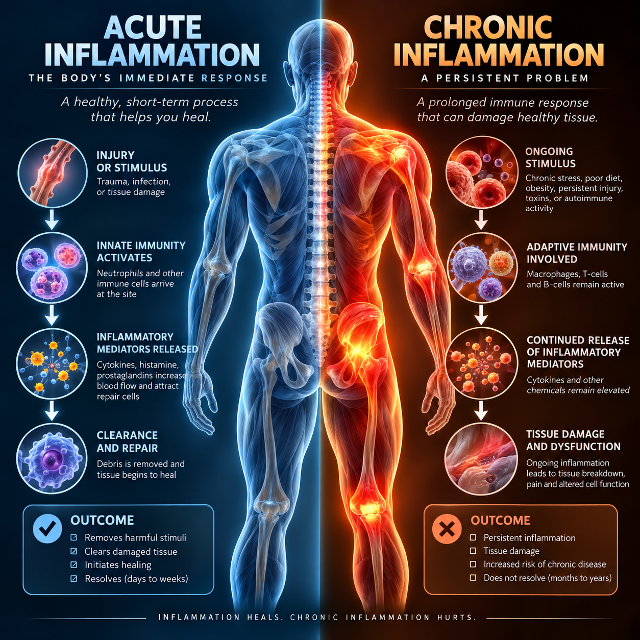
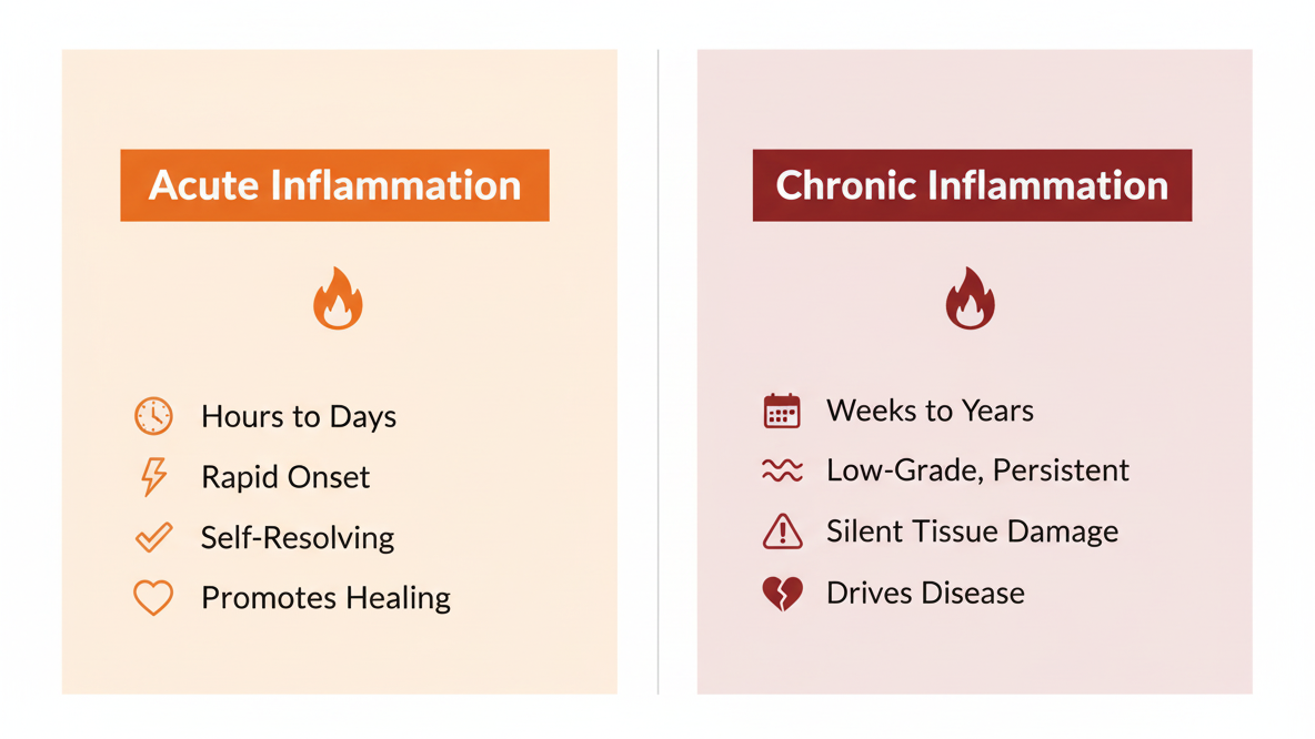
Acute vs Chronic Inflammation
Inflammation is your body’s repair signal; acute inflammation is short‑lived and essential for healing after injury, while chronic, low‑grade inflammation can delay recovery, worsen pain, and undermine the strength and resilience you’re trying to rebuild in rehab.
The Biology of Inflammation
Inflammation is not a single event — it is a coordinated cascade of molecular and cellular responses orchestrated by the innate immune system. When tissue is damaged or a pathogen is detected, resident immune cells release signaling proteins called cytokines — including interleukin-1β (IL-1β), interleukin-6 (IL-6), and tumor necrosis factor-alpha (TNF-α) — that amplify the immune response and recruit additional immune cells to the site. These signals activate the transcription factor NF-κB, a master regulator of inflammation that drives the expression of dozens of pro-inflammatory genes. (Medzhitov R. Nature, 2008)
Prostaglandins and leukotrienes, synthesized by COX (cyclooxygenase) and LOX (lipoxygenase) enzymes, mediate the classic signs of inflammation: heat, redness, swelling, and pain. These lipid mediators increase vascular permeability and sensitize nociceptors — which is why inflamed tissue hurts. NSAIDs like ibuprofen work precisely by inhibiting COX enzymes, blocking prostaglandin synthesis.
Critically, inflammation is designed to resolve. Once the threat is neutralized, the body produces specialized pro-resolving mediators (SPMs) — including lipoxins, resolvins, and protectins — derived largely from omega-3 fatty acids EPA and DHA. These molecules actively switch off the inflammatory response, clear debris, and initiate tissue repair. Failure of this resolution phase — not just excess activation — is increasingly recognized as central to chronic inflammatory disease. (Serhan CN. Nature Reviews Immunology, 2014)
When Inflammation Heals
Acute inflammation is one of the body’s most essential survival mechanisms. In response to injury or infection, the vascular response rapidly increases blood flow to the affected area, bringing neutrophils and macrophages that clear pathogens, remove debris, and release growth factors that initiate repair. This process is time-limited, self-resolving, and absolutely necessary — without it, wounds would not heal, infections would go unchecked, and muscle adaptation to exercise would not occur. (Rock KL et al. Nature Reviews Immunology, 2010)
Exercise-induced muscle damage is a well-studied example of beneficial acute inflammation. The micro-tears created by resistance training trigger an inflammatory response that recruits satellite cells, stimulates protein synthesis, and drives hypertrophy. Attempts to aggressively suppress post-exercise inflammation with NSAIDs or high-dose antioxidants have actually been shown to blunt these adaptations — a reminder that acute inflammation, when it resolves properly, is a feature, not a bug.
When Inflammation Harms
The problem arises when inflammation becomes chronic and low-grade — a persistent, smoldering state that lacks the clear trigger and resolution arc of acute inflammation. Chronic inflammation operates silently for years, driving tissue damage across multiple organ systems without producing the obvious signs of heat, redness, or swelling. It is now recognized as a foundational driver of most major chronic diseases of modern life. (Furman D et al. Nature Medicine, 2019)
In metabolic disease, chronic inflammation is both a cause and consequence. Excess visceral adipose tissue functions as an endocrine organ, secreting TNF-α, IL-6, and resistin — cytokines that impair insulin signaling and drive insulin resistance. In the cardiovascular system, inflammatory processes promote the formation and destabilization of atherosclerotic plaques. High-sensitivity C-reactive protein (hs-CRP), an acute-phase protein produced by the liver in response to IL-6, has emerged as an independent predictor of cardiovascular risk. (Hotamisligil GS. Nature, 2006; Libby P. Nature, 2002)
Neuroinflammation — inflammation within the central nervous system — is increasingly implicated in depression, cognitive decline, and neurodegenerative diseases including Alzheimer’s and Parkinson’s. The phenomenon of ‘inflammaging’ — the chronic, low-grade inflammatory state that accompanies aging — is associated with accelerated biological aging, sarcopenia, frailty, and reduced resilience to stress. (Furman D et al. Nature Medicine, 2019)
Lifestyle Factors That Drive Inflammation
Diet Quality
Ultra-processed foods, refined carbohydrates, and diets high in omega-6 fatty acids relative to omega-3s promote a pro-inflammatory environment. The modern Western diet has an estimated omega-6 to omega-3 ratio of 15–20:1, far above the evolutionary norm of approximately 4:1. This imbalance skews eicosanoid production toward pro-inflammatory pathways. (Simopoulos AP. Nutrients, 2016; Spreadbury I. Diabetes Metab Syndr Obes, 2012)
Physical Inactivity
Sedentary behavior is independently associated with elevated IL-6, CRP, and TNF-α — even after controlling for body composition. Skeletal muscle, when contracting regularly, releases anti-inflammatory myokines including IL-6 (in its exercise-induced, anti-inflammatory role) and IL-15 that modulate systemic immune function. (Petersen AM & Pedersen BK. J Appl Physiol, 2005)
Sleep Deprivation
Even short-term sleep restriction activates NF-κB signaling and elevates CRP and IL-6. Chronic sleep insufficiency (less than 6 hours per night) is associated with a 40–60% increase in systemic inflammatory markers compared to those sleeping 7–8 hours. (Irwin MR et al. Sleep, 2016)
Chronic Psychological Stress
Prolonged psychological stress dysregulates the HPA axis, leading to glucocorticoid resistance in immune cells — paradoxically making them less responsive to cortisol’s anti-inflammatory signals. This results in elevated pro-inflammatory cytokines and increased NF-κB activity. (Slavich GM & Irwin MR. Psychological Bulletin, 2014)
Visceral Adiposity
Visceral fat — the metabolically active fat surrounding abdominal organs — is a primary driver of systemic inflammation. Unlike subcutaneous fat, visceral adipocytes and their associated macrophages secrete substantial quantities of TNF-α and IL-6, creating a chronic pro-inflammatory milieu. (Wellen KE & Hotamisligil GS. J Clin Invest, 2005)
Gut Dysbiosis
Disruption of the gut microbiome impairs intestinal barrier integrity, allowing lipopolysaccharide (LPS) — a component of gram-negative bacterial cell walls — to translocate into systemic circulation. This ‘metabolic endotoxemia’ triggers low-grade inflammatory responses throughout the body and has been linked to insulin resistance, obesity, and non-alcoholic fatty liver disease. (Cani PD et al. Diabetes, 2008)
Lifestyle Strategies to Reduce Inflammation
The same modifiable lifestyle factors that drive inflammation can, when addressed, substantially reduce it. The evidence base for lifestyle-driven reductions in inflammatory biomarkers is robust and growing.
Anti-Inflammatory Diet
The Mediterranean dietary pattern — rich in vegetables, legumes, whole grains, olive oil, fish, and polyphenol-containing foods — is the most extensively studied anti-inflammatory diet. A landmark randomized controlled trial demonstrated that the Mediterranean diet reduced hs-CRP by 37% and IL-6 by 32% compared to a prudent control diet. (Esposito K et al. JAMA, 2004) Prioritizing omega-3 rich foods (fatty fish, walnuts, flaxseed) and minimizing refined carbohydrates and processed seed oils provides the most direct dietary influence on inflammatory mediators.
Regular Exercise
Habitual moderate-intensity exercise is one of the most powerful anti-inflammatory interventions available. Regular physical activity reduces TNF-α, CRP, and IL-6 at rest, and increases anti-inflammatory IL-10. Importantly, while a single acute bout of intense exercise transiently raises IL-6, the chronic adaptation to regular training is unambiguously anti-inflammatory. (Gleeson M et al. Nature Reviews Immunology, 2011)
Sleep Optimization
Prioritizing 7–9 hours of quality sleep per night is associated with the lowest inflammatory marker profiles across population studies. Sleep hygiene interventions — consistent sleep timing, dark and cool sleep environments, limiting blue light exposure — reduce NF-κB activation and normalize cytokine rhythms. (Irwin MR. Nature Reviews Immunology, 2019)
Stress Reduction
Mindfulness-based stress reduction (MBSR) has demonstrated significant reductions in IL-6 and CRP in randomized controlled trials. Other evidence-supported approaches include cognitive behavioral therapy (CBT), nature exposure, and social connection — all of which modulate HPA axis activity and downstream inflammatory signaling. (Black DS & Irwin MR. Ann NY Acad Sci, 2016)
Weight Management
Even modest weight loss of 5–10% of body weight produces significant reductions in CRP, IL-6, and TNF-α — effects largely attributable to reductions in visceral adipose tissue. The anti-inflammatory benefits of weight loss are independent of the method used to achieve it, whether through dietary restriction, exercise, or both. (Nicklas BJ et al. Arterioscler Thromb Vasc Biol, 2004)
Time-Restricted Eating
Intermittent fasting and time-restricted eating (TRE) protocols have demonstrated reductions in oxidative stress markers, CRP, and IL-6 independent of weight loss. TRE may exert anti-inflammatory effects through autophagy induction, circadian rhythm entrainment, and improvements in gut microbiome composition. (Moro T et al. J Transl Med, 2016)
Measuring Inflammation: Common Biomarkers
Several blood markers can help quantify the inflammatory burden — though none are perfectly specific, and individual results always require clinical interpretation. These reference ranges reflect population-level data and are provided for educational context only.
High-Sensitivity CRP (hs-CRP): The most widely used inflammatory biomarker in clinical practice. Values <1.0 mg/L indicate low cardiovascular risk; 1.0–3.0 mg/L moderate risk; >3.0 mg/L high risk. Values >10 mg/L typically indicate acute infection or injury rather than chronic inflammation.
Erythrocyte Sedimentation Rate (ESR): A non-specific marker of systemic inflammation. Useful for tracking disease activity in inflammatory conditions but less precise than hs-CRP.
Ferritin: An iron-storage protein that also functions as an acute-phase reactant. Elevated ferritin in the absence of iron deficiency often signals inflammatory or metabolic stress.
Homocysteine: An amino acid metabolite associated with cardiovascular inflammation and endothelial dysfunction. Elevated levels are linked to B-vitamin deficiency and increased cardiovascular risk.
Omega-6:Omega-3 Ratio: A functional marker of the body’s inflammatory balance. An ideal ratio is below 4:1; the typical Western diet produces ratios of 15–20:1. Can be assessed via specialized fatty acid panels.
The Resolution Phase: Inflammation Must Actively Resolve
A paradigm shift in inflammation research over the past two decades has been the recognition that resolution of inflammation is not merely the passive waning of pro-inflammatory signals — it is an active, programmed biological process. The failure to resolve, rather than simply excess initial activation, is now understood as a key driver of chronic inflammatory disease. (Serhan CN & Savill J. Nature Immunology, 2005)
Specialized pro-resolving mediators (SPMs) — including lipoxins, resolvins (derived from EPA and DHA), and protectins — are the molecular orchestrators of resolution. They halt neutrophil recruitment, promote macrophage-mediated clearance of cellular debris, and stimulate tissue regeneration. Dietary omega-3 fatty acids are the primary substrate for SPM synthesis, which is one mechanistic explanation for why omega-3 supplementation consistently demonstrates anti-inflammatory effects in clinical trials. (Calder PC. Biochim Biophys Acta, 2015)
References & Sources
- Furman D, et al. Chronic inflammation in the etiology of disease across the life span. Nature Medicine. 2019;25:1822–1832.
- Fullerton JN & Gilroy DW. Resolution of inflammation: a new therapeutic frontier. Nature Reviews Drug Discovery. 2016;15(8):551–567.
- Serhan CN. Pro-resolving lipid mediators are leads for resolution physiology. Nature. 2014;510(7503):92–101.
- Calder PC. Omega-3 fatty acids and inflammatory processes. Nutrients. 2010;2(3):355–374.
- Daily JW, et al. Efficacy of turmeric extracts and curcumin for alleviating the symptoms of joint arthritis. Journal of Medicinal Food. 2016;19(8):717–729.
- Meier-Ewert HK, et al. Effect of sleep loss on C-Reactive protein, an inflammatory marker of cardiovascular risk. Journal of the American College of Cardiology. 2004;43(4):678–683.
- Besedovsky L, et al. Sleep and immune function. Pflügers Archiv – European Journal of Physiology. 2012;463(1):121–137.
- Hotamisligil GS. Inflammation, metaflammation and immunometabolic disorders. Nature. 2017;542(7640):177–185.
- Tilg H & Moschen AR. Adipocytokines: mediators linking adipose tissue, inflammation and immunity. Nature Reviews Immunology. 2006;6(10):772–783.
- Konkel JE & Maruyama T. Gut-inflamed: how the microbiome regulates intestinal inflammation. Frontiers in Immunology. 2022;13:906258.
- Kabat-Zinn J. Full Catastrophe Living: Using the Wisdom of Your Body and Mind to Face Stress, Pain, and Illness. Delacorte Press. 1990.
- Pedersen BK & Saltin B. Exercise as medicine — evidence for prescribing exercise as therapy in 26 different chronic diseases. Scandinavian Journal of Medicine & Science in Sports. 2015;25(S3):1–72.
 Dr. Austin Fontenot